Dr. Zhenchao Dong and his coworkers at the International Center for Quantum Design of Functional Materials (ICQD) at Hefei National Laboratory for Physical Sciences at the Microscale in University of Science and Technology of China (USTC) has successfully visualized coherent intermolecular dipole-dipole coupling in real space using STM-based electroluminescence techniques. The work was published online in Nature on March 31, and the referees of Nature commented, “The fact that such effects could be directly imaged at the level of individual molecule is truly fascinating.” “I think this is a unique and exciting new way of studying dipole-dipole interactions between molecules with high significance for a broad community.”
A dipole describes the separation of positive and negative charges. A pair of electric charges of equal magnitude but opposite sign and separated by some small distance, for example, forms the simplest dipole. Dipole-dipole interaction between molecules, or coherent intermolecular dipole-dipole coupling, underlies important energy-transfer and optical processes in a wide range of scenarios, such as molecular interaction, photo synthesis light-harvesting and quantum optics. However, it is highly challenging to visualize coherent dipole coupling in real space at single molecular level owing to the diffraction limit in conventional optics.
As an old Chinese saying goes, one has to sharpen one’s tools first to get the job done. Scanning tunneling microscope (STM), with the very tiny metal tip used for position scanning, is a device that allows for imaging and manipulation of molecules at the atomic level. In 2010 the team developed a method to generate anomalous hot electro-luminescence for porphyrin molecules confined inside a plasmonic nanocavity in an STM, which can be applied to ultrahigh-resolution spectroscopy and sensing. Using this STM-induced electroluminescence method, the team successfully mapped the spatial distribution of the excitonic coupling in well-defined arrangements of a few zinc-phthalocyanine molecules.
In the research, molecules on the substrate were innovatively “pushed together” under STM techniques and imaged via sub-nanometer resolved single-molecule electroluminescence techniques. The luminescence patterns obtained from the dimer reveal the local optical response of the system and its dependence on the relative orientation and phase of the transition dipoles of the individual molecules in the dimer. Furthermore, enhanced ‘single-molecule’ superradiance was observed for in-line arrangementsup to four zinc-phthalocyanine molecules. “The findings we report clearly demonstrate that it is possible to visualize excitonic coupling in real space with sub-nanometre resolution”, says Dr. Zhenchao Dong, the group leader, “This capability should enable a greater understanding and rational engineering of light-harvesting structures and quantum light sources.” Yet the experimental approach may go much further and open up new fronts for the study of molecular interaction and energy transfer.
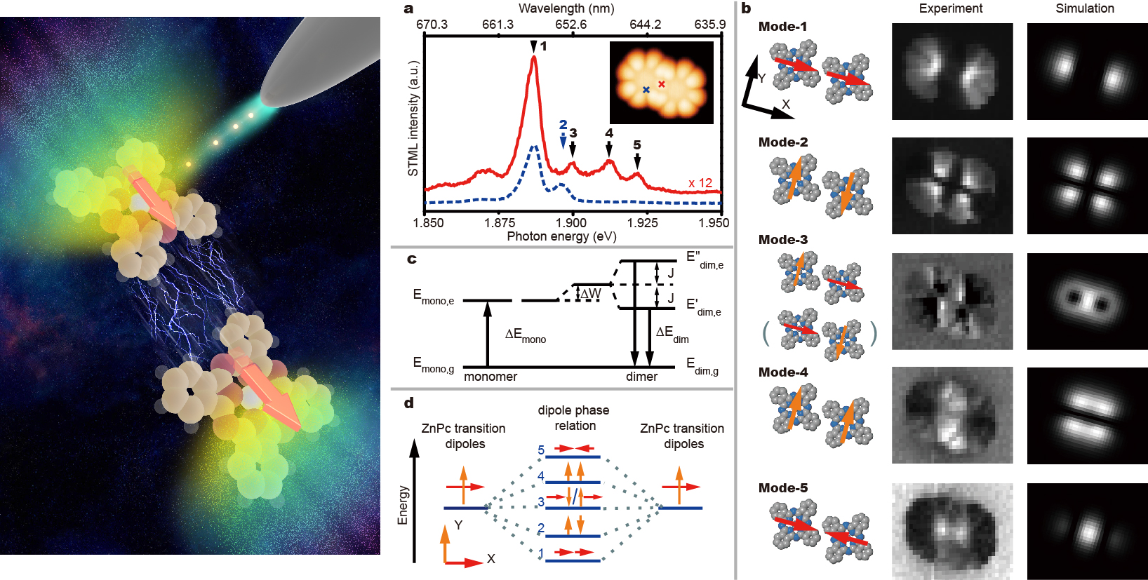
(Left) Artistic rendering of the experimental scheme (Right) The spatial distribution of the excitonic coupling of zinc-phthalocyanine dimers in real space mapped by STM-based electroluminescence at sub nanometer resolution.(Left provided by WANG Guoyan and SUN Daping, Right provided by DONG’s group)
Link of the publication: http://www.nature.com/nature/journal/v531/n7596/full/nature17428.html
From: http://en.ustc.edu.cn/highlight/201604/t20160405_240065.html